

International Journal of Infection 2026; 10(1) January-April: 5-9
ARTICLE
ROLE OF SERINE PROTEASES EXPRESSED IN IMMUNE CELLS AGAINST VIRAL INFECTIONS
Felaco M, De Lutiis MA, Grilli A. Role of serine proteases expressed in immune cells against viral infections. International Journal of Infection. 2026;10(1):5-9.
M. Felaco*, M.A. De Lutiis and A. Grilli
Department of Biomorphology, Faculty of Medicine and Surgery, Università degli Studi G. d’Annunzio, Chieti, Italy.
*Correspondence to:
Mario Felaco,
Department of Biomorphology,
Faculty of Medicine and Surgery,
Università degli Studi G. d’Annunzio,
Chieti, Italy.
e-mail: mfelaco@unich.it
| Received: 09 January, 2026 Accepted: 30 March, 2026  |
ISSN 3103-6678 [online] Copyright 2026 © by Biolife-publisher This publication and/or article is for individual use only and may not be further reproduced without written permission from the copyright holder. Unauthorized reproduction may result in financial and other penalties. Disclosure: all authors report no conflicts of interest relevant to this article. |
ABSTRACT
Serine proteases are enzymes expressed in immune cells that are important in the body’s defence against infection where they perform a degrading function and actively participate in the regulation and amplification of the immune response. They contribute to the destruction of infected cells through cytotoxic mechanisms mediated by cytotoxic T lymphocytes and natural killer (NK) cells, promoting the induction of apoptosis. Furthermore, they modulate the inflammatory response by activating or inactivating cytokines and receptors involved in the recruitment and activation of other immune cells.
KEYWORDS: Serine protease, enzyme, immune cell, infection, inflammation
INTRODUCTION
Serine proteases are expressed by several immune cells, such as CD8+ T cells, natural killer (NK) cells, and innate lymphocytes, and are capable of inducing cell death in virus-infected cells and tumors (1). Their name comes from the fact that they contain the amino acid serine in their active site, which is essential for their catalytic activity. Serine proteases play a fundamental role in the body’s defences.
Neutrophils and mast cells (MCs) release serine proteases, causing extracellular matrix degradation, cytokine activation, and tissue damage. Neutrophils are short-lived phagocytic immune cells that produce serine proteases such as elastase, proteinase 3, and cathepsin G, to destroy pathogenic microorganisms and modulate inflammation (2). MCs are immune cells involved in the innate and adaptive immune response. During the innate immune response, MCs release tryptase and chymase, which are key proteases in allergies and inflammation (3).
Serine proteases also play a crucial role in regulating inflammation and can act both as direct activators of inflammatory mediators and as indirect mediators, although many serine proteases are produced as inactive zymogens (4). Thrombin, complement (C1r, C1s), and the kallikrein-kinin system are involved in coagulation (5). Serine proteases activate complement molecules C3 and C5, which generate the anaphylatoxins C3a and C5a, which cause chemotaxis, increased permeability, and MC degranulation (6). Serine proteases also act on the kallikrein–kinin system, producing bradykinin, which mediates pain, edema, and vasodilation.
Serine proteases release sequestered growth factors and act on the extracellular matrix, causing the degradation of collagen, elastin, and proteoglycans (7). An active protease cleaves and activates other proteases, causing rapid amplification of inflammation. Humans have four protease-activated receptors (PARs): PAR-1, 2, 3, and 4 (8). Activation of PAR receptors by serine proteases is one of the main mechanisms of inflammation (9). The protease cleaves the extracellular domain of the receptor, exposing a ligand that activates the receptor. PAR receptors belong to the family of G-protein-coupled receptors (GPCRs) and are activated by proteases that cleave a portion of the receptor, exposing an internal ligand that activates it (10). Activation of G-proteins leads to an increase in intracellular calcium, which in turn, activates NF-kB and MAPK. These effects lead to the production of pro-inflammatory proteins such as IL-1, IL-6, and TNF, as well as adhesion molecules (11). Monocyte-macrophages are important cells for antigen presentation and the inflammatory response and can also express some serine proteases involved in regulating the immune response and tissue remodelling (12).
DISCUSSION
Serine proteases play an important role in protein degradation, both through peptide cleavage and in digestion, regulation, and cell signaling (13). They are characterized by a reactive serine in the active site. Trypsin, chymotrypsin, elastase, and thrombin are all serine proteases, which belong to the same enzyme family and share the same catalytic mechanism, but perform different biological functions (14). The function of these enzymes is to cut proteins into small peptides.
Serine proteases are enzymes that use a serine residue in the active site to cleave peptide bonds and participate in the destruction of microorganisms through various molecular mechanisms, both direct and indirect (15). All serine proteases share a catalytic triad that uses trace minerals at low doses to stimulate the body’s enzymatic and metabolic processes. Histidine activates serine, making it nucleophilic, and serine attacks the carbonyl carbon of the peptide bond of the substrate, forming a tetrahedral intermediate with cleavage of the peptide bond (16,17). This allows the formation of an acyl enzyme, a covalent intermediate that cleaves the protein. One portion of the protein is released, while the other remains bound to the acyl-enzyme intermediate. A water molecule that has been activated by histidine then enters, attacks the acyl intermediate, releases the second peptide fragment, and the enzyme returns to its initial state (18). The result is that one protein is formed into two peptides.
In the cellular machinery of protein degradation, serine functions on many cell types. For example, in the digestive tract, dietary proteins are broken down into amino acids (19). Some proteases regulate the activation of other proteins, apoptosis, and the immune response. Each serine protease recognizes specific sequences, such as trypsin, which cleaves after basic amino acids (Lys, Arg); chymotrypsin, which cleaves after aromatic residues; and elastase, which acts in the small intestine to degrade dietary proteins, facilitating their digestion and absorption, to create small residues (20).
Serine proteases are produced as inactive zymogens, e.g., trypsinogen-trypsin, and are regulated by protease inhibitors (21). The main difference between the various serine proteases is not the mechanism, but the substrate specificity and biological function. For example, trypsin cleaves after basic amino acids (lysine, arginine), has a protein-digesting function in the intestine, and activates other zymogens with a primary role (22). Chymotrypsin cleaves after aromatic amino acids (phenylalanine, tyrosine, tryptophan) and has a digestive function (23).
Effect of Serine Proteases on Microorganisms
Viruses are obligate intracellular organisms that use the host cell to reproduce and infect cells by binding to specific membrane receptors. They enter through membrane fusion or endocytosis. Inside the host cell, the virus can release its genetic material, which can be DNA or RNA, and uses cellular metabolism to replicate its genome. Infected cells are recognized and destroyed by immune cells (24).
Serine proteases are important enzymes of the immune system that help kill bacteria, viruses, fungi, and parasites. NK cells and cytotoxic T cells recognize tumor and virus-infected target cells as foreign and kill them through the release of perforin, which creates pores in the target cell membrane, and granzymes that enter the cell through the pores produced by perforin (25). Granzymes activate caspases, damage DNA, destroy proteins vital to the target cell, and cause apoptosis (26). In humans, there are 5 different types of granzymes: granzyme A, B, H, K, and M, and granzyme B is faster and more active in causing damage to target cells by rapidly activating apoptosis (27,28).
Serine proteases activate complement proteins, leading to the formation of the membrane attack complex (MAC) (29). This leads to pathogen entrapment and direct lysis, resulting in the activation of coagulation factors, the formation of pro-inflammatory molecules, and the recruitment of immune cells (Fig.1). Serine proteases can cleave microorganism surface proteins, alter their membranes, and destroy harmful molecules. These serine effects facilitate cellular phagocytosis, kill the microorganism, and enhance the immune system (30).
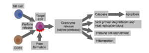
Fig. 1. Serine proteases are expressed by immune cells that play a crucial role in the defense against viral infections. Granzymes released by CD8+ cytotoxic T lymphocytes and natural killer (NK) cells are essential for eliminating infected cells. After recognizing the viral antigen, cytotoxic T lymphocytes release granules containing perforin and granzymes. Perforin facilitates the formation of pores in the target cell membrane, allowing the granzymes to enter. Once inside, these serine proteases activate apoptotic cascades through caspase activation, leading to programmed cell death and limiting the spread of the virus. Serine protease mediates inflammation and cellular recruitment.
CONCLUSIONS
Serine proteases are a family of highly versatile enzymes, with both effector and regulatory functions. They act by using a serine residue in the active site to cleave peptide bonds and participate in the destruction of microorganisms both directly and indirectly. Furthermore, they participate in the regulation of adaptive immunity, thus contributing to the response against viruses.
Therefore, serine proteases represent an important weapon for controlling viral infections, but they can potentially have pathological and inflammatory effects. A thorough understanding of their mechanisms of action could open new therapeutic avenues, both to enhance the antiviral response and to limit inflammation.
Conflict of interest
The authors declare that they have no conflict of interest.
REFERENCES
- Donado CA, Theisen E, Zhang F, et al. Granzyme K activates the entire complement cascade. Nature. 2025;641(8061):211-221. doi:https://doi.org/10.1038/s41586-025-08713-9
- Kettritz R. Neutral serine proteases of neutrophils. Immunological Reviews. 2016;273(1):232-248. doi:https://doi.org/10.1111/imr.12441
- Mukai K, Tsai M, Starkl P, Marichal T, Galli SJ. IgE and mast cells in host defense against parasites and venoms. Seminars in Immunopathology. 2016;38(5):581-603. doi:https://doi.org/10.1007/s00281-016-0565-1
- Santos FX, Arroyo C, Garcı́aI, et al. Role of mast cells in the pathogenesis of postburn inflammatory response: reactive oxygen species as mast cell stimulators. Burns. 2000;26(2):145-147. doi:https://doi.org/10.1016/s0305-4179(99)00021-2
- Irmscher S, Döring N, Halder LD, et al. Kallikrein Cleaves C3 and Activates Complement. Journal of Innate Immunity. 2017;10(2):94-105. doi:https://doi.org/10.1159/000484257
- Deng S, Liao J, Li H, et al. Streptococcus suis subtilisin-like serine proteases SspA-1 and SspA-2 interplay with complement C3a and C5a to facilitate bacterial immune evasion and infection. Virulence. 2024;15(1). doi:https://doi.org/10.1080/21505594.2023.2301246
- Youssef ME, Abdel-Reheim MA, Morsy MA, et al. Ameliorative Effect of Dabigatran on CFA-Induced Rheumatoid Arthritis via Modulating Kallikrein-Kinin System in Rats. International Journal of Molecular Sciences. 2022;23(18):10297. doi:https://doi.org/10.3390/ijms231810297
- Pawar N, Buzza MS, Antalis TM. Membrane-Anchored Serine Proteases and Protease-Activated Receptor-2–Mediated Signaling: Co-Conspirators in Cancer Progression. Cancer Research. 2019;79(2):301-310. doi:https://doi.org/10.1158/0008-5472.can-18-1745
- Bunnett N. Protease-Activated Receptors: How Proteases Signal to Cells to Cause Inflammation and Pain. Seminars in Thrombosis and Hemostasis. 2006;32(S 1):039-048. doi:https://doi.org/10.1055/s-2006-939553
- Hara T, Sata M, Fukuda D. Emerging roles of protease-activated receptors in cardiometabolic disorders. Journal of Cardiology. 2023;81(4):337-346. doi:https://doi.org/10.1016/j.jjcc.2022.09.013
- Nieuwenhuizen L, Falkenburg WJJ, Schutgens REG, et al. Stimulation of Naïve Monocytes and PBMCs with Coagulation Proteases Results in Thrombin‐Mediated and PAR‐1‐Dependent Cytokine Release and Cell Proliferation in PBMCs Only. Scandinavian Journal of Immunology. 2013;77(5):339-349. doi:https://doi.org/10.1111/sji.12033
- Moriuchi H, Moriuchi M, Fauci AS. Cathepsin G, a Neutrophil-Derived Serine Protease, Increases Susceptibility of Macrophages to Acute Human Immunodeficiency Virus Type 1 Infection. Journal of Virology. 2000;74(15):6849-6855. doi:https://doi.org/10.1128/jvi.74.15.6849-6855.2000
- Fedoriuk M, Stefanenko M, Bohovyk R, et al. Serine proteases and protease-activated receptors signaling in the kidney. American Journal of Physiology-Cell Physiology. 2025;329(1):C107-C117. doi:https://doi.org/10.1152/ajpcell.00143.2025
- Penny GS, Dyckes DF. Interaction of dansylated peptidyl chloromethanes with trypsin, chymotrypsin, elastase, and thrombin. Biochemistry. 1980;19(13):2888-2894. doi:https://doi.org/10.1021/bi00554a011
- Hooper NM. Proteases: a primer. Hooper NM, ed. Essays in Biochemistry. 2002;38:1-8. doi:https://doi.org/10.1042/bse0380001
- Ekici ÖD, Paetzel M, Dalbey RE. Unconventional serine proteases: Variations on the catalytic Ser/His/Asp triad configuration. Protein Science. 2008;17(12):2023-2037. doi:https://doi.org/10.1110/ps.035436.108
- Gololobov G, Tramontano A, Paul S. Nucleophilic Proteolytic Antibodies. Applied Biochemistry and Biotechnology. 2000;83(1-3):221-232. doi:https://doi.org/10.1385/abab:83:1-3:221
- Sreenivasan U, Axelsen PH. Buried water in homologous serine proteases. Biochemistry. 1992;31(51):12785-12791. doi:https://doi.org/10.1021/bi00166a011
- Camilleri M, Sellin JH, Barrett KE. Pathophysiology, Evaluation, and Management of Chronic Watery Diarrhea. Gastroenterology. 2017;152(3):515-532.e2. doi:https://doi.org/10.1053/j.gastro.2016.10.014
- Shi YJ, Huang CH, Lee YC, Wang LJ, Chiou JT, Chang LS. Naja atra cardiotoxins enhance the protease activity of chymotrypsin. International Journal of Biological Macromolecules. 2019;136:512-520. doi:https://doi.org/10.1016/j.ijbiomac.2019.06.066
- Fernández I, Saunders N, Stéphane Duquerroy, et al. Structural basis of TMPRSS2 zymogen activation and recognition by the HKU1 seasonal coronavirus. Cell. 2024;187(16):4246-4260.e16. doi:https://doi.org/10.1016/j.cell.2024.06.007
- Sun M, Wei S, Li Y, et al. Utilizing a Negative Enrichment Strategy to Profile Protein Methylation, Leveraging the Orthogonality of LysargiNase and Trypsin. Molecular & Cellular Proteomics. 2025;24(7):100970. doi:https://doi.org/10.1016/j.mcpro.2025.100970
- Kong WW, Yan YL, Hou CP, et al. A novel digestive protease chymotrypsin-like serine contributes to anti-BmNPV activity in silkworm (Bombyx mori). Developmental & Comparative Immunology. 2025;162:105301. doi:https://doi.org/10.1016/j.dci.2024.105301
- Hoffmann M, Kleine-Weber H, Schroeder S, et al. SARS-CoV-2 Cell Entry Depends on ACE2 and TMPRSS2 and Is Blocked by a Clinically Proven Protease Inhibitor. Cell. 2020;181(2):271-280. doi:https://doi.org/10.1016/j.cell.2020.02.052
- Voskoboinik I, Whisstock JC, Trapani JA. Perforin and granzymes: function, dysfunction and human pathology. Nature Reviews Immunology. 2015;15(6):388-400. doi:https://doi.org/10.1038/nri3839
- Zhang Z, Zhang Y, Xia S, et al. Gasdermin E suppresses tumor growth by activating anti-tumor immunity. Nature. 2020;579(7799):415-420. doi:https://doi.org/10.1038/s41586-020-2071-9
- Janiszewski T, Kołt S, Kaiserman D, et al. Noninvasive optical detection of granzyme B from natural killer cells with enzyme-activated fluorogenic probes. Journal of Biological Chemistry. 2020;295(28):9567-9582. doi:https://doi.org/10.1074/jbc.ra120.013204
- Miyashita H, Kurzrock R, Bevins NJ, et al. T-cell priming transcriptomic markers: implications of immunome heterogeneity for precision immunotherapy. npj Genomic Medicine. 2023;8(1). doi:https://doi.org/10.1038/s41525-023-00359-8
- Garred P, Tenner AJ, Mollnes TE. Therapeutic Targeting of the Complement System: From Rare Diseases to Pandemics. Pharmacological Reviews. 2021;73(2):792-827. doi:https://doi.org/10.1124/pharmrev.120.000072
- Fuchs TA, Abed U, Goosmann C, et al. Novel cell death program leads to neutrophil extracellular traps. The Journal of Cell Biology. 2007;176(2):231-241. doi:https://doi.org/10.1083/jcb.200606027