

International Journal of Infection 2026; 10(3) September-December: 59-62
THE TRANSCRIPTION FACTOR c-Fos REGULATES THE IMMUNE RESPONSE AGAINST BACTERIA AND VIRUSES
Stuard S, de Iuliis A. The transcription factor cFos regulates the immune response against bacteria and viruses. International Journal of Infection. 2026;10(3):59-62.
S. Stuard1 and A. de Iuliis2*
1 Global Medical Office – Clinical Affairs, CoE Clinical & Therapeutic Governance, Fresenius Medical Care, Bad Homburg, Germany;
2 Clinical Pathology, Azienda Sanitaria Locale 2, Atessa Hospital, Lanciano-Vasto, Chieti, Italy.
*Correspondence to:
Antonio de Iuliis, MD,
Clinical Pathology,
Azienda Sanitaria Locale 2,
Atessa Hospital, Lanciano-Vasto,
Chieti, Italy.
e-mail: Antoniodeiuliis@alice.it
| Received: 20 July, 2026 Accepted: 04 September, 2026  |
ISSN 3103-6678 [online] Copyright 2026 © by Biolife Publisher This publication and/or article is for individual use only and may not be further reproduced without written permission from the copyright holder. Unauthorized reproduction may result in financial and other penalties. Disclosure: all authors report no conflicts of interest relevant to this article. |
ABSTRACT
The FOS gene is an immediate-response proto-oncogene that encodes the c-Fos protein, a transcription factor that, by forming the activator protein-1 (AP-1) complex with c-Jun, regulates genes involved in proliferation, differentiation, inflammation, and stress response, and is also an important marker of neuronal activation. In addition to being involved in tumors and epilepsy, FOS plays a key role in the immune response and neuronal activation, being also induced during infections through the activation of Toll-like receptors (TLRs) and mitogen-activated protein kinase (MAPK) signaling pathways. TLR9 activation by microbial DNA induces the MAPK pathway, which activates c-Fos/AP-1, promoting the expression of pro-inflammatory cytokines involved in the immune response against bacteria and viruses. However, some viruses exploit or alter these pathways to promote their own replication or evade the immune system, while AP-1 often cooperates with NF-κB and nuclear factor of activated T cells (NFAT) for full activation of cytokine transcription. Overall, the FOS/c-Fos–AP-1 signaling axis is a central regulator linking pathogen recognition to inflammatory, antiviral, and neuronal responses, while its dysregulation or viral manipulation contributes to disease pathogenesis.
KEYWORDS: c-Fos, transcription factor, infection, immune response, bacteria, virus
INTRODUCTION
The FOS gene encodes mRNA FOS, leading to the protein c-Fos, which is rapidly expressed following neuronal stimulation or other cellular signals. c-Fos functions as a transcription factor that regulates the expression of downstream target genes. It forms the activator protein-1 (AP-1) complex through dimerization with proteins such as c-Jun, allowing regulation of genes involved in cellular processes including stress responses, proliferation, differentiation, inflammation, survival, and apoptosis (1,2).
c-Fos is widely used as a molecular marker of neuronal activation. Following exposure to a strong stimulus, insult, antigen, or pharmacological agent, c-Fos expression increases rapidly, typically becoming detectable within approximately 30-60 minutes and reaching peak levels within a few hours (1,3). Because c-Fos expression occurs in activated neurons, it can be used to identify neuronal populations recruited during specific stimuli or behavioral states. Detection methods include c-Fos gene expression analysis and immunohistochemistry that allow researchers to map activated neural circuits and investigate processes such as anxiety, depression, pain, substance addiction, memory formation, sleep, and wakefulness (4).
DISCUSSION
The FOS gene, also known as an immediate early gene (IEG) because of its rapid response to cellular activation, is a proto-oncogene that mediates certain pathologies such as tumors and epilepsy. In some tumors, FOS is altered or persistently increased (2). c-Fos activation by inflammatory signals involves the generation of interleukin (IL)-2 and tumor necrosis factor (TNF) which participate in the activation of T lymphocytes and act against infections (5). In the central nervous system (CNS), c-Fos is a marker of neurons. It indicates brain areas activated after stress and pain and is an important marker of neuronal circuits (1). c-Fos can be activated during an infection when a bacterium or virus infects the body (6).
In general, microorganisms are recognized through pattern recognition receptors (PRRs), particularly Toll-like receptor (TLR)4, which recognizes the lipopolysaccharide (LPS) of Gram-negative bacteria (7). In viral infections, TLR3 recognizes double-stranded RNA and TLR9 recognizes DNA rich in unmethylated cytosine–phosphate–guanine (CpG) motifs (6). CpG is a DNA sequence in which a cytosine (C) is followed by a guanine (G) on the same strand, with the phosphate group (p) of the sugar-phosphate backbone between the two nucleotides. In mammalian DNA, many CpG sites are methylated (cytosine can become 5-methylcytosine); while in bacteria and many viruses, CpG motifs are common and generally unmethylated. TLR9 uses these reactions to recognize microbial DNA and activate the innate immune response. Activated TLR triggers a biochemical cascade of intracellular signaling that includes myeloid differentiation primary response 88 (MyD88), IL-1 receptor-associated kinase (IRAK), TNF receptor-associated factor 6 (TRAF6), transforming growth factor-β-activated kinase 1 (TAK1), and mitogen-activated protein kinases (MAPKs), including extracellular signal-regulated kinase (ERK), tyrosine kinase non-receptor (TNK), and p38 (8).
MAPK and c-Fos activation
c-Fos, MAPK activation, and the immune response against bacteria and viruses are interconnected concepts in the signaling pathway of several TLRs, including TLR9, and the mechanism follows pathogen recognition. In endosomes, TLR9 recognizes DNA with unmethylated CpG motifs from bacteria and viruses, resulting in activation of the TLR9 pathway (9). This pathway recruits MyD88, which activates several kinases, including MAPK, ERK, c-Jun N-terminal kinase (JNK), and p38. c-Fos activation occurs when MAPKs induce the expression of the transcription factor c-Fos, which associates with c-Jun to form the AP-1 complex (2). AP-1 is involved in the immune response by promoting the transcription of genes encoding the pro-inflammatory cytokines TNF, IL-6, IL-1β, chemokines, and other molecules involved in the immune response against bacteria and viruses (10) (Fig.1).
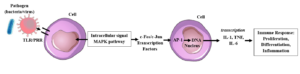
Fig. 1. Toll-like receptor (TLR)9 activation initiates a biochemical cascade involving transcription factors such as NF-κB and activator protein-1 (AP-1), inducing the transcription of genes and the secretion of cytokines involved in the immune response against bacteria and viruses.
The reaction between c-Fos, MAPKs, and microorganisms activates infectious mechanisms and an immune response. TLRs detect viral and bacterial proteins, a recognition that activates intracellular signaling pathways, including MAPKs, the main ones being ERK, JNK, and p38 (11).
In bacterial infections, activation of the MAPK/c-Fos pathway primarily promotes the inflammatory response, which is important for eliminating bacteria (10). In viral infections, however, in addition to promoting inflammation, they also induce antiviral genes (5).
Several viruses have evolved strategies to modulate or exploit the MAPK and c-Fos pathways to their advantage, promoting replication or limiting the immune response (12). In addition to bacteria, viruses also activate MAPKs to increase c-Fos and promote their own replication, to use AP-1 to transcribe viral genes, or to inhibit the aforementioned pathways to evade the immune system (13). For example, influenza A virus activates p38 and the endoplasmic reticulum, and herpes simplex virus exploits AP-1 during the replication cycle, while severe acute respiratory syndrome coronavirus 2 (SARS-CoV-2) can alter MAPK pathways, contributing to the generation of pro-inflammatory cytokines (14,15). Furthermore, AP-1 activation alone can often be insufficient to produce robust cytokine generation (2). Therefore, the transcription of many cytokines requires the cooperation of AP-1 with other transcription factors, especially NF-κB and, in some cells and situations, with nuclear factor of activated T cells (NFAT), as occurs, for example, with the expression of IL-2 in T lymphocytes (5) (Fig.2).

Fig. 2. Microbial DNA, including bacterial and viral nucleic acids, activates Toll-like receptors (TLRs), such as TLR9, triggering the mitogen-activated protein kinase (MAPK) signaling pathway and induction of the immediate-early gene FOS. The resulting c-Fos protein heterodimerizes c-Jun to form the activator protein-1 (AP-1) transcription factor, which regulates genes involved in inflammation, antiviral responses, proliferation, differentiation, stress responses, and neuronal activation. AP-1 can cooperate with NF-κB and nuclear factor of activated T cells (NFAT) to promote the transcription of pro-inflammatory cytokines and other immune-response genes. Some viruses can exploit or modulate these signaling pathways to enhance their replication or evade host immune responses. Dysregulation of the FOS/c-Fos–AP-1 axis may contribute to pathological processes, including infection-associated inflammation, tumorigenesis, and neuronal dysfunction.
CONCLUSIONS
The FOS gene is an immediate-early proto-oncogene that encodes c-Fos, a transcription factor that forms the AP-1 complex with c-Jun to regulate genes involved in cell proliferation, differentiation, inflammation, stress responses, neuronal activation, and immunity. During bacterial and viral infections, activation of TLRs (particularly TLR9) triggers MAPK signaling, leading to c-Fos/AP-1 activation and the expression of pro-inflammatory and antiviral genes, although some viruses can manipulate these pathways to enhance replication or evade immune responses; full cytokine expression often requires cooperation between AP-1, NF-κB, and NFAT.
Conflict of interest
The authors declare that they have no conflict of interest.
REFERENCES
- Hoffman GE, Smith MS, Verbalis JG. c-Fos and Related Immediate Early Gene Products as Markers of Activity in Neuroendocrine Systems. Frontiers in Neuroendocrinology. 1993;14(3):173-213. doi:10.1006/frne.1993.1006
- Eferl R, Wagner EF. AP-1: a double-edged sword in tumorigenesis. Nature Reviews Cancer. 2003;3(11):859-868. doi:10.1038/nrc1209
- Sundquist SJ, Nisenbaum LK. Fast Fos: rapid protocols for single- and double-labeling c-Fos immunohistochemistry in fresh frozen brain sections. Journal of Neuroscience Methods. 2005;141(1):9-20. doi:10.1016/j.jneumeth.2004.05.007
- Gao YJ, Ji RR. c-Fos or pERK, Which is a Better Marker for Neuronal Activation and Central Sensitization After Noxious Stimulation and Tissue Injury? The Open Pain Journal. 2009;2(1):11-17. doi:10.2174/1876386300902010011
- Bird L. Staying in character. Nature Reviews Immunology. 2007;7(2):86-87. doi:10.1038/nri2031
- Chaudhuri J, Alt FW. Class-switch recombination: interplay of transcription, DNA deamination and DNA repair. Nature Reviews Immunology. 2004;4(7):541-552. doi:10.1038/nri1395
- Miller EK. The prefrontal cortex and cognitive control. Nature reviews Neuroscience. 2000;1(1):59-65. doi:10.1038/35036228
- Li X, Jiang S, Tapping RI. Toll-like receptor signaling in cell proliferation and survival. Cytokine. 2010;49(1):1-9. doi:10.1016/j.cyto.2009.08.010
- Dalpke A, Frank J, Peter M, Heeg K. Activation of Toll-Like Receptor 9 by DNA from Different Bacterial Species. Infection and Immunity. 2006;74(2):940-946. doi:10.1128/iai.74.2.940-946.2006
- Chervonsky AV. Influence of microbial environment on autoimmunity. Nature Immunology. 2009;11(1):28-35. doi:10.1038/ni.1801
- Salauddin MD, Bhattacharyya D, Samanta I, et al. Role of TLRs as signaling cascades to combat infectious diseases: a review. Cellular and Molecular Life Sciences. 2025;82(1). doi:10.1007/s00018-025-05631-x
- Chathuranga K, Weerawardhana A, Dodantenna N, Lee JS. Regulation of antiviral innate immune signaling and viral evasion following viral genome sensing. Experimental & Molecular Medicine. 2021;53(11):1647-1668. doi:10.1038/s12276-021-00691-y
- Mohanta TK, Sharma N, Arina P, Defilippi P. Molecular Insights into the MAPK Cascade during Viral Infection: Potential Crosstalk between HCQ and HCQ Analogues. BioMed Research International. 2020;2020(1):8827752-8827752. doi:10.1155/2020/8827752
- Keyaerts E, Vijgen L, Pannecouque C, et al. Plant lectins are potent inhibitors of coronaviruses by interfering with two targets in the viral replication cycle. Antiviral Research. 2007;75(3):179-187. doi:10.1016/j.antiviral.2007.03.003
- Bošnjak B, Stein SC, Willenzon S, et al. Low serum neutralizing anti-SARS-CoV-2 S antibody levels in mildly affected COVID-19 convalescent patients revealed by two different detection methods. Cellular & Molecular Immunology. 2020;18(4):936-944. doi:10.1038/s41423-020-00573-9