

European Journal of Neurodegenerative Diseases 2024; 13(1) January-April AHEAD OF PRINT
BIOLOGICAL EFFECTS OF SUBSTANCE P IN THE BRAIN
I. Robuffo*
CNR Section of Chieti, 66100 Chieti, Italy.
*Correspondence to:
Iole Robuffo, MD,
CNR Section of Chieti,
66100 Chieti, Italy.
e-mail: iole.robuffo@cnr.it
| Received: 13 February, 2024 Accepted: 29 February, 2024 |
2974-6345 (2024) Copyright © by BIOLIFE This publication and/or article is for individual use only and may not be further reproduced without written permission from the copyright holder. Unauthorized reproduction may result in financial and other penalties. Disclosure: all authors report no conflicts of interest relevant to this article. |
ABSTRACT
Substance P (SP) is a non-cholinergic neuropeptide produced at different levels in different cell types, including mast cells (MCs) and neurons. SP is involved in the regulation of many central nervous system (CNS) disorders such as anxiety, stress, mood disorders, and neurogenesis, and functions such as synapse growth, dendritic formation, respiration, neurotoxicity, nociception and pain. This neuropeptide produces inflammation after binding to its natural killer (NK) cell receptor. MCs and neurons possess the NK1 receptor and are activated by SP to produce inflammatory molecules such as cytokines and chemokines. In this article, we report that SP not only induces the generation of pro-inflammatory proteins, but also acts synergistically with cytokines such as IL-33 to enhance the inflammatory process.
KEYWORDS: substance P, brain, CNS, immunology, inflammation
INTRODUCTION
Substance P (SP) was discovered in 1931 by V. Euler and Gaddum who found that extracts from brain and intestinal tissues had hypotensive and spasmogenic activity. Subsequently, a protein was extracted from these tissues which was called SP. Further studies highlighted that SP is a neuropeptide member of the tachykinin (TAC) family that is present in mammals and distinguished in three subgroups: TAC1, 3, and 4. SP is a non-cholinergic neurotransmitter that is present at different concentrations in different areas of the central nervous system (CNS) (1). For example, SP is found at higher concentrations in the dorsal spinal roots, while it is lower in the ventral roots (2). Levels of SP are elevated in the grey matter, midbrain, postrema area, nuclei, and medullary fibers (3). However, SP concentrations in various brain areas could be species-specific and therefore vary in the various experimental animals used (4).
SP is involved in the regulation of anxiety, stress, mood disorders, neurogenesis, the growth of synapses, dendritic formation, respiration, neurotoxicity, and nociception and pain (5). When SP is injected into the third ventricle in experimental animals, it stimulates respiration and causes a slight increase in blood pressure (6).
DISCUSSION
Substance P (SP) in inflammation
SP is a protein made up of 11 amino acids that is produced by several cell types, including neurons and immune cells (7). SP acts by binding to its G protein-coupled neurokinin receptors (NKR), which include NK1R, NK2R, and NK3R (8). From these receptors, NK1R appears to have greater affinity for SP. SP binds the NK1R receptor in immune cells, causing an immune response, including the reaction towards microbes (9). In addition, SP has been observed to mediate tissue homeostasis and wound healing (9).
SP can mediate neurogenic inflammation and can be released after stimulation of the sensory nerves (10,11). This neuropeptide is biologically active in endothelial cells and smooth muscle cells and increases vascular permeability with the consequent leakage of plasma and the formation of edema (12,13). SP also activates intracellular adhesion molecules (ICAMs) as well as vascular cell adhesion molecules (VCAMs) in vascular epithelial cells (14,15). Mast cells (MCs) treated in vitro with SP generate more VEGF than untreated samples (16,17). The action of SP on the vessels causes an increase in vascularization with inflammatory cells crossing the tissues (18,19). SP works by binding to its NK1 receptor and mediates itching, which can be inhibited by antagonist drugs that block NK1 (20,21) (Fig.1).
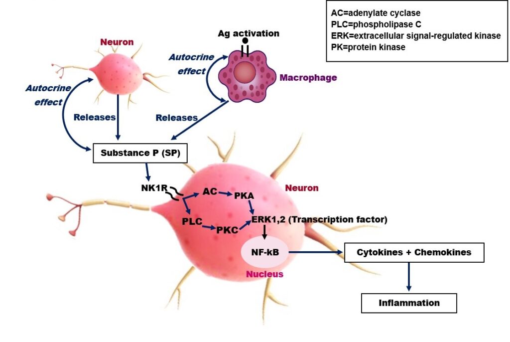
Fig. 1. A schematic reproduction depicting Substance P (SP) secretion by neurons and macrophages.
Substance P and mast cells (MCs)
MCs are immune cells derived from bone marrow that migrate and mature in tissues throughout the human body, which mediate innate and adaptive immunity (22,23). In addition to expressing the FceRI receptor, MCs show other ligands on their surface, including those of neuropeptides such as SP (24,25).
It has been reported that IL-33 significantly increases the ability of SP to stimulate MCs to secrete VEGF, tumor necrosis factor (TNF), and IL-1β, selectively without tryptase granule release (26,27) In their interesting article, Theoharides et al. provide much information on SP-mediated inflammation in relation to IL-33 (26).
In an in vitro study, the treatment of a line of MCs (LAD2) with SP plus the cytokine IL-33 achieved a synergistic effect on the stimulation of the potent pro-inflammatory cytokine IL-1β, compared to the administration of cells treated with IL-33 alone (28). These results demonstrate that SP is not only a potent pro-inflammatory molecule, but that it is also able to synergistically enhance other inflammatory cytokines such as IL-33 (29,30). In addition, the authors reported that SP alone has a stimulatory effect on IL-1β production in MCs only at high concentrations (1µM), while at lower concentrations (0.01–0.1 µM) there was no effect. These effects were also confirmed by IL-1β gene expression which demonstrated an active participation of SP in inflammatory effects induced by IL-1β. However, these reactions did not affect the inflammasome protein (NLRP3 or ASC) levels. Blocking the NK1 receptor on MCs suppressed the secretion of IL-1β. In this study, it was evident that SP and its receptor NK1 were involved in the induction of IL-1β. The authors concluded that in combination with IL-33, SP synergistically induces IL-1β secretion from MCs.
CONCLUSIONS
In conclusion, SP that is produced by different cells, including neurons and MCs, induces cytokines and chemokines that mediate many neurological disorders. In addition, by binding its receptor NK1, SP can act synergistically with certain cytokines, such as IL-33, and induce inflammation.
Conflict of interest
The author declares that they have no conflict of interest.
REFERENCES
- Wang XF, Ge TT, Fan J, Yang W, Cui RJ. The role of substance P in epilepsy and seizure disorders. Oncotarget. 2017;8(44):78225-78233. doi:https://doi.org/10.18632/oncotarget.20606
- Akagi H, Konishi S, Otsuka M, Yanagisawa M. The role of substance P as a neurotransmitter in the reflexes of slow time courses in the neonatal rat spinal cord. British Journal of Pharmacology. 1985;84(3):663-673. doi:https://doi.org/10.1111/j.1476-5381.1985.tb16148.x
- Raoof M, Soofiabadi S, Abbasnejad M, Kooshki R, Esmaeili‐Mahani S, Mansoori M. Activation of orexin‐1 receptors in the ventrolateral periaqueductal grey matter (vlPAG) modulates pulpal nociception and the induction of substance P in vlPAG and trigeminal nucleus caudalis. International Endodontic Journal. 2018;52(3):318-328. doi:https://doi.org/10.1111/iej.13007
- Denis P, Fardin V, Nordmann JP, et al. Localization and characterization of substance P binding sites in rat and rabbit eyes. Investigative ophthalmology & visual science. 1991;32(6):1894-1902.
- Ebner K, Singewald N. The role of substance P in stress and anxiety responses. Amino Acids. 2006;31(3):251-272. doi:https://doi.org/10.1007/s00726-006-0335-9
- Ciccocioppo R, Polidori C, Pompei P, De Caro G, Massi M. Inhibition of isotonic sodium chloride intake in the rat by selective tachykinin agonists. Pharmacology, biochemistry and behavior. 1994;47(3):609-615. doi:https://doi.org/10.1016/0091-3057(94)90166-x
- Cottrell GS, Hooper NM, Turner AJ. Cloning, Expression, and Characterization of Human Cytosolic Aminopeptidase P: A Single Manganese(II)-Dependent Enzyme. Biochemistry. 2000;39(49):15121-15128. doi:https://doi.org/10.1021/bi001585c
- Regoli D, Drapeau G, Dion S, D’Orléans-Juste P. Pharmacological receptors for substance P and neurokinins. Life Sciences. 1987;40(2):109-117. doi:https://doi.org/10.1016/0024-3205(87)90349-3
- Suvas S. Role of Substance P Neuropeptide in Inflammation, Wound Healing, and Tissue Homeostasis. The Journal of Immunology. 2017;199(5):1543-1552. doi:https://doi.org/10.4049/jimmunol.1601751
- Choi JE, Di Nardo A. Skin Neurogenic inflammation. Seminars in immunopathology. 2018;40(3):249-259. doi:https://doi.org/10.1007/s00281-018-0675-z
- Marek-Jozefowicz L, Nedoszytko B, Grochocka M, et al. Molecular Mechanisms of Neurogenic Inflammation of the Skin. International Journal of Molecular Sciences. 2023;24(5):5001. doi:https://doi.org/10.3390/ijms24055001
- Bolton TB, Clapp LH. Endothelial-dependent relaxant actions of carbachol and substance P in arterial smooth muscle. British Journal of Pharmacology. 1986;87(4):713-723. doi:https://doi.org/10.1111/j.1476-5381.1986.tb14589.x
- Budel S, Schuster A, Stergiopoulos N, Meister JJ, Bény JL. Role of smooth muscle cells on endothelial cell cytosolic free calcium in porcine coronary arteries. American Journal of Physiology-heart and Circulatory Physiology. 2001;281(3):H1156-H1162. doi:https://doi.org/10.1152/ajpheart.2001.281.3.h1156
- Quinlan KL, Naik SM, Cannon G, et al. Substance P activates coincident NF-AT- and NF-kappa B-dependent adhesion molecule gene expression in microvascular endothelial cells through intracellular calcium mobilization. Journal of immunology. 1999;163(10):5656-5665.
- Münzel T, Heitzer T, Harrison DG. The physiology and pathophysiology of the nitric oxide/superoxide system. Herz. 1997;22(3):158-172. doi:https://doi.org/10.1007/bf03044353
- Theoharides TC, Zhang B, Kempuraj D, et al. IL-33 augments substance P–induced VEGF secretion from human mast cells and is increased in psoriatic skin. Proceedings of the National Academy of Sciences. 2010;107(9):4448-4453. doi:https://doi.org/10.1073/pnas.1000803107
- Sismanopoulos N, Delivanis DA, Mavrommati D, Hatziagelaki E, Conti P, Theoharides TC. Do mast cells link obesity and asthma? Allergy. 2012;68(1):8-15. doi:https://doi.org/10.1111/all.12043
- O’Connor TM, O’Connell J, O’Brien DI, Goode T, Bredin CP, Shanahan F. The role of substance P in inflammatory disease. Journal of Cellular Physiology. 2004;201(2):167-180. doi:https://doi.org/10.1002/jcp.20061
- Castellani ML, Galzio RJ, P. Felaco, et al. VEGF, substance P and stress, new aspects: a revisited study. Journal of biological regulators & homeostatic agents. 2010;24(3):229-237.
- Vander Does A, Ju T, Mohsin N, Chopra D, Yosipovitch G. How to get rid of itching. Pharmacology & Therapeutics. 2023;243:108355. doi:https://doi.org/10.1016/j.pharmthera.2023.108355
- Chung BY, Kim HB, Jung MJ, et al. Post-Burn Pruritus. International Journal of Molecular Sciences. 2020;21(11):3880. doi:https://doi.org/10.3390/ijms21113880
- Theoharides TC, Donelan JM, Papadopoulou N, Cao J, Kempuraj D, Conti P. Mast cells as targets of corticotropin-releasing factor and related peptides. Trends in Pharmacological Sciences. 2004;25(11):563-568. doi:https://doi.org/10.1016/j.tips.2004.09.007
- Theoharides TC, Petra AI, Taracanova A, Panagiotidou S, Conti P. Targeting IL-33 in Autoimmunity and Inflammation. Journal of Pharmacology and Experimental Therapeutics. 2015;354(1):24-31. doi:https://doi.org/10.1124/jpet.114.222505
- Lauritano D, Mastrangelo F, D’Ovidio C, et al. Activation of Mast Cells by Neuropeptides: The Role of Pro-Inflammatory and Anti-Inflammatory Cytokines. International Journal of Molecular Sciences. 2023;24(5):4811. doi:https://doi.org/10.3390/ijms24054811
- Conti P, Lauritano D, Caraffa A, et al. Mast Cells Mediate Rheumatoid Arthritis- Inhibitory Role of IL-37. Critical Reviews in Immunology. 2019;39(4):267-274. doi:https://doi.org/10.1615/critrevimmunol.2020033176
- Theoharides TC. Effect of Stress on Neuroimmune Processes. Clinical Therapeutics. 2020;42(6):1007-1014. doi:https://doi.org/10.1016/j.clinthera.2020.05.002
- Castellani ML, Vecchiet J, Salini V, et al. Stimulation of CCL2 (MCP-1) and CCL2 mRNA by substance P in LAD2 human mast cells. Translational Research. 2009;154(1):27-33. doi:https://doi.org/10.1016/j.trsl.2009.03.006
- Taracanova A, Alevizos M, Karagkouni A, et al. SP and IL-33 together markedly enhance TNF synthesis and secretion from human mast cells mediated by the interaction of their receptors. Proceedings of the National Academy of Sciences of the United States of America. 2017;114(20):E4002-E4009. doi:https://doi.org/10.1073/pnas.1524845114
- Castellani Ml, Ciampoli C, Felaco M, et al. Neuropeptide Substance P induces mRNA expression and secretion of CXCL8 chemokine, and HDC in human umbilical cord blood mast cells. Clinical and investigative medicine. 2008;31(6):362-362. doi:https://doi.org/10.25011/cim.v31i6.4923
- Nicoletti M, Neri G, Maccauro G, et al. Impact of Neuropeptide Substance P an Inflammatory Compound on Arachidonic Acid Compound Generation. International Journal of Immunopathology and Pharmacology. 2012;25(4):849-857. doi:https://doi.org/10.1177/039463201202500403