

European Journal of Neurodegenerative Diseases 2026; 15(3) September-December: 60-64
p38 MAPK SIGNAL PATHWAY ACTIVATION IN PARKINSON’S DISEASE
M.J. Ross1* and F. Gaudelli2
1 Dermatology, Greenwich Medical Spa, Greenwich, USA;
2 Department of Clinical and Experimental Medicine, University of Foggia, Foggia, Italy.
*Correspondence to:
Mitch J. Ross, MD,
Greenwich Medical Spa,
Greenwich, USA.
e-mail: mitchjross@gmail.com
| Received: 03 August, 2026 Accepted: 22 September, 2026  |
ISSN 2279-5855 print / 3103-7364 [online] Copyright 2026 © by Biolife Publisher This publication and/or article is for individual use only and may not be further reproduced without written permission from the copyright holder. Unauthorized reproduction may result in financial and other penalties. Disclosure: All authors report no conflicts of interest relevant to this article. |
ABSTRACT
p38 mitogen-activated protein kinase (MAPK) is a key intracellular signalling pathway of the MAPK family that mediates cellular stress, inflammation, infection, and immune responses through the activation of transcription factors and the production of cytokines. In contrast, ERK1/2 primarily regulates cell growth and proliferation through the RAS–RAF–MEK–ERK pathway, whereas c-Jun N-terminal Kinase (JNK) is mainly involved in cellular stress, inflammation, and apoptosis. Among the p38 MAPK isoforms, p38-α (MAPK14) is the most extensively studied and is activated by stress-related stimuli through the MAP3K–MKK3/6–p38 pathway, contributing to inflammation, oxidative stress, neuronal death, and mitochondrial dysfunction in neurological diseases such as Parkinson’s disease (PD). PD is a multifactorial neurodegenerative disorder characterized by the progressive and selective loss of dopaminergic neurons in the substantia nigra pars compacta. α-synuclein aggregation, mitochondrial dysfunction, oxidative stress, and damage-associated molecular pattern (DAMP)-mediated microglial activation contribute to p38 MAPK–mediated neuroinflammation and neuronal death. The resulting reduction in striatal dopamine disrupts the balance between the D1-mediated direct and D2-mediated indirect basal ganglia pathways, leading to reduced thalamocortical activation and contributing to motor symptoms such as bradykinesia and akinesia. Moreover, α-synuclein accumulation, mitochondrial dysfunction, and oxidative stress promote microglial activation and stimulate the MKK3/6–p38 MAPK pathway, increasing the production of pro-inflammatory cytokines and promoting neuronal vulnerability and apoptosis. This creates a self-reinforcing cycle in which neuroinflammation, oxidative stress, mitochondrial dysfunction, and p38 MAPK activation contribute to further dopaminergic neuron degeneration, highlighting p38 MAPK as a potential link between molecular stress responses and neurodegeneration in PD.
KEYWORDS: Parkinson’s disease, p38, MAPK, signal pathway activation, neurodegeneration
INTRODUCTION
p38 mitogen-activated protein kinase (MAPK) is one of the MAPK subfamilies that is particularly involved in responses to cellular stress, inflammation, and cytokine generation (1). p38 MAPK is an important intracellular signalling pathway implicated in the innate immune response in the recognition of infectious microorganisms, leading to inflammation, although it also participates in the adaptive response (2). Microorganisms activate pathogen-associated molecular patterns (PAMPs) and damage-associated molecular patterns (DAMPs), the pattern recognition receptors (PRRs) activating MAPK3K, inducing MKK 3/6, and p38 MAPK, inducing transcription factors and the generation of cytokines (1). Activating stimuli can include lipopolysaccharide (LPS) from Gram-negative bacteria which binds to TLR4, viral RNA which binds to TLR3/7/8 and RIG-I/MDA5, bacterial lipoproteins which bind to TLR2, and reactive oxygen species (ROS) that cause tissue damage (3).
p38 MAPK acts intracellularly in the response to stress, inflammation, cell damage, and infection (4). ERK1/2, which is involved in cell proliferation, growth, and differentiation, also belongs to the MAPK family. While ERK1/2 is a mitogenic growth factor, c-Jun N-terminal kinase (JNK) is involved in cellular stress, inflammation, and apoptosis. For example, ERK1/2 can be activated by epidermal growth factor (EGF), causing proliferation, while JNK can be activated by UV, causing c-Jun activation in response to stress (1). Stimulus activates renin-angiotensin system (RAS), which activates MEK1/2 and ERK1/2, leading to proliferation and growth; stress, on the other hand, activates MKKK and MKK4/7, which activate Jun and c-Jun/AP-1, which trigger the stress response and apoptosis (1). Thus, ERK activates growth signals, while JNK activates stress signals (5).
The main p38 MAPK isoforms are p38-α (MAPK14), p38-b, p38-g, and p38-d. Among these, the best known and most studied is p38-α from immune cells. It is activated by various stimuli that activate MAP3K and MKK3/6, which stimulate and induce p38 and the phosphorylation of substrates (6). Activation of p38 MAPK pathways is linked to neuroinflammation, neuronal death, oxidative stress, and mitochondrial dysfunction, which occur in neurological diseases such as Parkinson’s disease (PD) (7,8).
PARKINSON’S DISEASE AND p38 MAPK
PD is the second most common neurodegenerative disease and the fastest growing worldwide. It is characterized by tremor, rigidity, slowness, and difficulty with movement. The disease does not exclusively affect the elderly, as it has been noted that one in four patients presents with the onset before age 50. Cases have doubled in the last 15 years, and a further increase is estimated in the next 20 years (9,10).
PD is a multifactorial disease characterized by the selective death of dopaminergic neurons located in the substantia nigra pars compacta (SNpc), with alterations in mitochondria, proteases, autophagy, and α-synuclein (8). Monomeric α-synuclein transforms into oligomers that activate fibrils and thus Lewy bodies. Oligomers are toxic and harmful and can alter cell membranes and the ubiquitin-proteasome system. Additionally, they can promote oxidative stress, interfere with vesicle trafficking, and alter mitochondrial function (11,12). Oxidative stress can damage dopaminergic neurons, triggering a chain reaction that compromises the structure and function of the brain (13). Usually, nervous tissue is particularly vulnerable to this phenomenon because it consumes enormous amounts of oxygen and is rich in oxidation-sensitive fats (14).
In PD, p38 MAPK links cellular stress, neuroinflammation, and dopaminergic neuronal death (15,16). Moreover, aggregated α-synuclein leads to mitochondrial dysfunction and activation, increased ROS, and neuronal alteration (17). Exposure to DAMPs activates microglia, which produce inflammatory mediators and cause neuroinflammation (17). Aggregated extracellular α-synuclein acts as a damage signal and activates microglia, which release inflammatory cytokines, mainly IL-1, TNF, and IL-6 (17,18). ATP released by cells can activate P2X7, leading to the release of HMGB1 from damaged cells and the activation of TLR4/RAGE (19). Mitochondrial DNA can activate innate immune pathways, such as cGAS-STING, and oxidative stress, thereby promoting the release and activation of damage signals (20). Thus, the dysfunctional neuron proves to be a source of danger signals and neuroinflammation (Fig.1) (20).
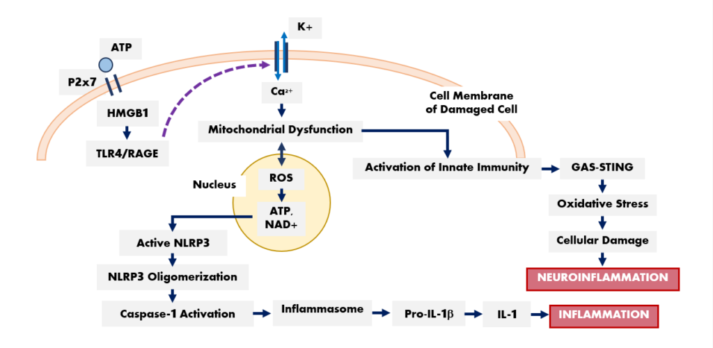
Fig. 1. ATP binds to the P2x7 receptor, which activates TLR4/RAGE signaling and the calcium–potassium ion pump, leading to mitochondrial dysfunction and subsequent activation of innate immunity and inflammation. ROS activate ATP and NAD+, promoting the formation of the NLRP3 inflammasome, caspase-1, and IL-1, thereby contributing to inflammation.
p38 MAPK AND THE DECREASE IN DOPAMINE IN PARKINSON’S DISEASE
The decrease in dopamine in PD is due to the degeneration of dopaminergic neurons in the SNpc with alterations in the basal ganglia, which is a crucial aspect of the disease (21). In PD, dopaminergic neurons undergo progressive degeneration with a decrease in dopamine levels in the striatum (22). The remaining neurons consist of α-synuclein aggregates or Lewy bodies (23). Dopamine is crucial because it modulates two important pathways: the direct pathway, mediated by D1 receptors, which increases activity and facilitates movement, and the indirect pathway, mediated by D2 receptors, which decreases activity and suppresses movement (24). Therefore, dopamine facilitates the initiation and execution of movement, but when it decreases, D1 stimulation occurs, activating the indirect pathway and resulting in reduced motor facilitation. Simultaneously, D2 decreases, which increases the indirect pathway to result in greater motor inhibition (24). These reactions decrease thalamocortical activity, resulting in decreased stimulation of the motor cortex, with bradykinesia/akinesia (24,25).
PD degeneration is multifactorial and is characterized by oxidative stress, impaired proteostasis/autophagy, and neurodegeneration (8,26). In neurodegeneration, various stimuli accumulate that can activate the MAPK cascade, particularly p38 (7). Oxidative stress, α-synuclein, and mitochondrial dysfunction mediate neuroinflammation with activation of MAP3K, MKK3/MKK6, and p38 MAPK, leading to activation of transcription factors and a pro-inflammatory response and cellular stress (apoptosis and neurodegeneration) (7,26). In nigrostriatal dopaminergic neurons, increased ROS and mitochondrial dysfunction can activate p38 MAPK, leading to microglia activation and the production of IL-1 and TNF, which can induce stimulation of the p38 pathway (8). The decrease in dopaminergic neurons creates neuroinflammation and an increase in p38 MAPK (7). p38 MAPK can phosphorylate several targets and modulate the expression of genes involved in neuroinflammation. Moreover, long-lasting p38 stimulation can cause neuronal vulnerability and apoptosis (26).
In PD, the interplay between p38 MAPK and dopamine links neuroinflammation, oxidative stress, mitochondrial dysfunction, and the death of dopaminergic neurons (27). Low dopamine levels, resulting from the loss of dopaminergic neurons, impair basal ganglia circuitry and contribute to motor symptoms (28). Neuronal death is driven by α-synuclein acting on mitochondria, triggering ROS production, microglial activation, cytokine release, and p38 MAPK signalling, in a series of self-reinforcing reactions that lead to neuronal damage (29). p38-α (MAPK14) is the most extensively studied isoform and is primarily activated by stress stimuli. It is triggered by ROS, TNF-α, or IL-1β, and its activation engages the MKK3/MKK6/p38 pathway, leading to the phosphorylation of nuclear and cytoplasmic substrates (7). p38 activation can modulate gene expression, mitochondrial function, inflammatory responses, and cell survival (26). Dopamine itself can contribute to oxidative stress; elevated cytosolic dopamine levels can undergo oxidation to generate ROS and quinone products, while its metabolic breakdown via monoamine oxidase (MAO) contributes to H₂O₂ production (8,30). Thus, damage signals can activate microglia and the p38 pathway, contributing to the production of pro-inflammatory cytokines that can damage dopaminergic neurons (17).
CONCLUSIONS
p38 MAPK appears to play a central role in the complex interplay between oxidative stress, mitochondrial dysfunction, α-synuclein accumulation, neuroinflammation, and dopaminergic neuronal death in PD. Its persistent activation may contribute to a self-sustaining cycle of microglial activation, pro-inflammatory cytokine production, and neuronal degeneration. Therefore, p38 MAPK may represent an important molecular link between cellular stress and neurodegeneration, as well as a potential therapeutic target for slowing disease progression and protecting dopaminergic neurons.
Conflict of interest
The authors declare that they have no conflict of interest.
REFERENCES
- Hu Y, Song J, Liu L, et al. Different microRNA alterations contribute to diverse outcomes following EV71 and CA16 infections: Insights from high-throughput sequencing in rhesus monkey peripheral blood mononuclear cells. The International Journal of Biochemistry & Cell Biology. 2016;81:20-31. doi:10.1016/j.biocel.2016.10.011
- Rambow F, Rogiers A, Marin-Bejar O, et al. Toward Minimal Residual Disease-Directed Therapy in Melanoma. Cell. 2018;174(4):843-855.e19. doi:10.1016/j.cell.2018.06.025
- Spencer SP, Fragiadakis GK, Sonnenburg JL. Pursuing human-relevant gut microbiota-immune interactions. Immunity. 2019;51(2):225-239. doi:10.1016/j.immuni.2019.08.002
- Coulthard LR, White DE, Jones DL, McDermott MF, Burchill SA. p38MAPK: stress responses from molecular mechanisms to therapeutics. Trends in Molecular Medicine. 2009;15(8):369-379. doi:10.1016/j.molmed.2009.06.005
- Lavoie H, Gagnon J, Therrien M. ERK signalling: a master regulator of cell behaviour, life and fate. Nature Reviews Molecular Cell Biology. 2020;21(10):607-632.
- Canovas B, Nebreda AR. Diversity and versatility of p38 kinase signalling in health and disease. Nature Reviews Molecular Cell Biology. 2021;22(5):346-366. doi:10.1038/s41580-020-00322-w
- Keeler JF, Pretsell DO, Robbins TW. Functional implications of dopamine D1 vs. D2 receptors: A ‘prepare and select’ model of the striatal direct vs. indirect pathways. Neuroscience. 2014;282:156-175. doi:10.1016/j.neuroscience.2014.07.021
- Rocha EM, De Miranda B, Sanders LH. Alpha-synuclein: Pathology, mitochondrial dysfunction and neuroinflammation in Parkinson’s disease. Neurobiology of Disease. 2018;109(0969-9961):249-257. doi:10.1016/j.nbd.2017.04.004
- Dorsey ER, Sherer T, Okun MS, Bloem BR. The Emerging Evidence of the Parkinson Pandemic. Journal of Parkinson’s Disease. 2018;8(S1):S3-S8. doi:10.3233/jpd-181474
- Riboldi GM, Frattini E, Monfrini E, Frucht SJ, Di Fonzo A. A Practical Approach to Early-Onset Parkinsonism. Journal of Parkinson’s Disease. 2022;12(1):1-26. doi:10.3233/jpd-212815
- Alam P, Bousset L, Melki R, Otzen DE. α‐synuclein oligomers and fibrils: a spectrum of species, a spectrum of toxicities. Journal of Neurochemistry. 2019;150(5):522-534. doi:10.1111/jnc.14808
- Ingelsson M. Alpha-Synuclein Oligomers-Neurotoxic Molecules in Parkinson’s Disease and Other Lewy Body Disorders. Frontiers in Neuroscience. 2016;10. doi:10.3389/fnins.2016.00408
- Esteves AR, Arduíno DM, Silva DFF, Oliveira CR, Cardoso SM. Mitochondrial Dysfunction: The Road to Alpha-Synuclein Oligomerization in PD. Parkinson’s Disease. 2011;2011:1-20. doi:10.4061/2011/693761
- Branco DM, Arduino DM, Esteves AR, et al. Cross-talk between mitochondria and proteasome in Parkinson’s disease pathogenesis. Frontiers in Aging Neuroscience. 2010 ;2:17. doi:10.3389/fnagi.2010.00017
- Jeohn GH, Cooper CL, Wilson B, et al. p38 MAP Kinase Is Involved in Lipopolysaccharide‐Induced Dopaminergic Neuronal Cell Death in Rat Mesencephalic Neuron‐Glia Cultures. Annals of the New York Academy of Sciences. 2002;962(1):332-346. doi:10.1111/j.1749-6632.2002.tb04078.x
- Shalabi MG, Al-kuraishy HM, Shokr MM, Batiha GE. Molecular crosstalk between MAPK signaling and neuroprotective pathways in Parkinson’s disease: from pathogenesis to therapeutic potential. Neuroscience. 2026;610:297-310. doi:10.1016/j.neuroscience.2026.06.026
- Zhang QS, Heng Y, Yuan YH, Chen NH. Pathological α-synuclein exacerbates the progression of Parkinson’s disease through microglial activation. Toxicology Letters. 2017;265:30-37. doi:10.1016/j.toxlet.2016.11.002
- Hoffmann A, Ettle B, Bruno A, et al. Alpha-synuclein activates BV2 microglia dependent on its aggregation state. Biochemical and Biophysical Research Communications. 2016;479(4):881-886. doi:10.1016/j.bbrc.2016.09.109
- Chen R, Kang R, Tang D. The mechanism of HMGB1 secretion and release. Experimental & Molecular Medicine. 2022;54(2):91-102. doi:10.1038/s12276-022-00736-w
- Chakraborty R, Maya S, Testa V, et al. α-Synuclein aggregates induce mitochondrial damage and trigger innate immunity to drive neuron–microglia communication. Nature Communications. 2026;17(1). doi:10.1038/s41467-026-73136-7
- Frosini D, Cosottini M, Volterrani D, Ceravolo R. Neuroimaging in Parkinson’s disease: focus on substantia nigra and nigro-striatal projection. Current Opinion in Neurology. 2017;30(4):416-426. doi:10.1097/wco.0000000000000463
- Heng N, Malek N, Lawton M, et al. Striatal Dopamine Loss in Early Parkinson’s Disease: Systematic Review and Novel Analysis of Dopamine Transporter Imaging. Movement Disorders Clinical Practice. 2023;10(4):539-546. doi:10.1002/mdc3.13687
- Choong CJ, Mochizuki H. Neuropathology of α‐synuclein in Parkinson’s disease. Neuropathology. 2022;42(2):93-103. doi:10.1111/neup.12812
- Gerfen CR. Segregation of D1 and D2 dopamine receptors in the striatal direct and indirect pathways: An historical perspective. Frontiers in Synaptic Neuroscience. 2023;14. doi:10.3389/fnsyn.2022.1002960
- Wichmann T, DeLong MR, Guridi J, Obeso JA. Milestones in research on the pathophysiology of Parkinson’s disease. Movement Disorders. 2011;26(6):1032-1041. doi:10.1002/mds.23695
- Patel VB, Zhabyeyev P, Chen X, et al. PI3Kα-regulated gelsolin activity is a critical determinant of cardiac cytoskeletal remodeling and heart disease. Nature communications. 2018;9(1). doi:10.1038/s41467-018-07812-8
- Chakrabarti S, Bisaglia M. Oxidative Stress and Neuroinflammation in Parkinson’s Disease: The Role of Dopamine Oxidation Products. Antioxidants. 2023;12(4):955. doi:10.3390/antiox12040955
- Zhou Z, Yi L, Wang Q, Lim TM, Tan EK. Role of dopamine in the pathophysiology of Parkinson’s disease. Translational Neurodegeneration. 2023;12(1). doi:10.1186/s40035-023-00378-6
- Hyemi Eo, Kim S, Jung UJ, Kim SR. Alpha-Synuclein and Microglia in Parkinson’s Disease: From Pathogenesis to Therapeutic Prospects. Journal of Clinical Medicine. 2024;13(23):7243-7243. doi:10.3390/jcm13237243
- Gendaszewska-Darmach E, Drzazga A, Koziołkiewicz M. Targeting GPCRs Activated by Fatty Acid-Derived Lipids in Type 2 Diabetes. Trends in Molecular Medicine. 2019;25(10):915-929. doi:10.1016/j.molmed.2019.07.003
