

European Journal of Neurodegenerative Diseases 2026; 15(3) September-December: 49-54
INDUCTION AND PRODUCTION OF CHEMOKINES IN THE CNS BY VIRUS-INFECTED CELLS
A. di Stadio1*, L. Neri2 and G. Neri3
1 Department of Life Sciences, Health and Health Professional, Link University of Rome, Rome, Italy;
2 ENT Specialist, ASL Lanciano, Vasto, Chieti;
3 Neurosciences and Imaging, University of Chieti-Pescara, Chieti, Italy.
*Correspondence to:
Dr. Arianna di Stadio,
Department of Life Sciences, Health and Health Professional,
Link University of Rome,
Rome, Italy.
e-mail: a.distadio@unilink.it
| Received: 20 July, 2026 Accepted: 10 September, 2026  |
ISSN 2279-5855 print / 3103-7364 [online] Copyright 2026 © by Biolife Publisher This publication and/or article is for individual use only and may not be further reproduced without written permission from the copyright holder. Unauthorized reproduction may result in financial and other penalties. Disclosure: All authors report no conflicts of interest relevant to this article. |
ABSTRACT
Chemokines are proteins that regulate cell migration, especially that of immune cells, which increase during infection and inflammation. They are activated by G protein-coupled receptors (GPCRs) and the Gi protein, to participate in various intracellular pathways involved in cytoskeletal reorganization and chemotaxis. In the central nervous system (CNS), chemokines contribute to neuroinflammation, while during infections, they coordinate leukocyte recruitment through integrin activation and the phases of rolling, adhesion, diapedesis, and chemotaxis, linking microorganism recognition to the inflammatory response. Cells in the brain recognize viral genetic material through innate immune receptors, activating pathways such as NF-κB, mitogen-activated protein kinase (MAPK), and AP-1, which induce the production of chemokines that attract specific immune cells and coordinate the antiviral response. CD8+ lymphocytes are activated by T-cell receptor (TCR)/major histocompatibility complex class I (MHC-I), costimulation, and cytokines, to proliferate and reach infected tissues thanks to chemokines, where they eliminate infected cells via perforin, granzymes, or FAS-FASL and produce interferon gamma (IFN-γ), promoting the antiviral response, the expression of MHC-I, and the activation of macrophages. Virus-infected cells in the brain produce type I IFNs and pro-inflammatory cytokines that induce chemokines such as CXCL10, CCL2, CXCL9, and CCL5. These chemokines attract lymphocytes, natural killer (NK) cells, and monocytes/macrophages to the CNS, contributing to infection control and neuroinflammation. In neurodegenerative diseases and during brain damage, chemokines activate microglia and astrocytes and can cause inflammation, oxidative stress, and alterations in the blood-brain barrier (BBB). Chemokines can induce neurodegeneration and neuronal damage, but in some cases, they also promote repair, neuronal survival, and neuroprotection, depending on the chemokine, receptor, cell type, and disease stage.
KEYWORDS: Chemokine, virus, infection, CNS, immune cell
INTRODUCTION
Chemokines are small proteins involved in cell migration and attraction that primarily affect immune cells (1) They are produced by various cells, including endothelial cells, immune cells, and fibroblasts (2). Neurotropic viruses that infect the central nervous system (CNS) can activate an immune response in which chemokines play a key role (3).
Viruses are recognized by the CNS and induce the production of cytokines and chemokines that recruit immune cells and cause neuroinflammation and cellular degeneration (3). In inflammatory processes, cell damage, and infections, chemokines increase their production and bind specifically to their receptors (2). It is known that CXC chemokines primarily attract neutrophils, whereas CC chemokines are chemotactic for lymphocytes and macrophages. Chemokines activate G proteins, causing a conformational change in the receptor. Activation of the Gai protein reduces the activity of adenylate cyclase and activates various intracellular pathways (1). The main signalling pathways activated by chemokines are PI3K/Akt, PLCb/Ca2+, Ra/MAPK, and the activation of Rho, Rac, and Cdc42, which are important for the cytoskeleton (1).
Chemokines are essential for cellular chemotaxis, resulting in the directional migration of the target cell toward the source of the chemokine. The cell forms a “leading edge” facing the generation of the chemokine and rearranges actin via Rac and Cdc42 and forms cellular protrusions (4). In addition, the cell establishes adhesion to the substrate, contracts the trailing edge via Rho/ROCK and myosin, advancing towards the gradient (4). Chemokines cause leukocyte recruitment in inflammatory processes by coordinating immune cell trafficking and intervening in both the cellular and humoral immune responses, where they guide immune cells circulating in the peripheral blood (5,6).
Chemokines are produced in inflamed tissue and presented on the endothelial surface. Neutrophils possess chemokine receptors and come into contact with selectins present on the endothelium. Chemokines act on G protein-coupled receptors (GPCRs) (1,2). In microbial infections, macrophages, fibroblasts, dendritic cells, endothelial cells, and others produce IL-1, TNF, IL-6, and other inflammatory cytokines, causing tissue damage (7,8) (Fig.1). In particular, chemokines bind to their receptors by activating the Gi protein which activates PI3K/PLC and mitogen-activated protein kinase (MAPK) (1). These reactions lead to an increase in intracellular Ca2+, PIP3, activation of AKT and Rac/Rho/Cdc42. Rac/Rho/Cdc42 are involved in the reorganization of the cytoskeleton, causing cell wall concentration and directional movement (4). In addition, chemokines transform leukocyte integrins, making them more high affinity (5,6) The integrins LFA-1 (CD11a/CD18) and Mac-1 (CD11bCD18) bind to ICAM-1/ICAM-2 molecules, allowing the rolling phase with subsequent activation, adhesion, diapedesis, and chemotaxis (6). In infections, chemokines are important because they link microorganism recognition to leukocyte migration to the site of inflammation (9).

Fig. 1. Neutrophils produce chemokines that bind endothelial cells through E-selectin which induces an immune response with the release of pro-inflammatory cytokines, leading to inflammation.
DISCUSSION
Cells in the brain, such as microglia, astrocytes, and endothelial cells, recognize viral RNA or DNA via innate immunity receptors (10). After a pathogenic virus infects tissue, immune cells rush to the area and recognize pathogen-associated molecular patterns (PAMPs) through pattern recognition receptors (PRRs) (11). Toll-like receptor (TLR) activation on the cell membrane stimulates NOD1/NOD2 in the cytoplasm and RIG-1/MDA5, which induces viral RNA and cGAS-STING, which binds DNA and leads to the production of inflammasomes with caspase-1 activation (10,11). After several reactions, TLR activation leads to the generation of NF-kB and, in parallel, TAK1 with activation of MAPK and AP-1 (9). These molecules enter the nucleus and increase chemokine transcription (9). Each specific chemokine binds to its receptor and recruits specific immune cells (12) (Table I).
Table I. Some chemokine ligands with their corresponding receptors.
| Ligand | Receptor |
| CXCL8 | CXCR1, 2 |
| CXCL11, 12 | CCR7 |
| CXCL16 | CXCR6 |
| CXCL13 | CXCR5 |
| CXCL12 | CXCR4 |
| CX3CL1-CCL26 | CX3CR1 |
| CXCL1, 5, 7 | ECRF3 |
| CCL2, 3, 4, 5 | US28 |
| CCL2,3,4,5 | UL12 |
| CCL3, 4, 5, 7, 14, 15, 16 | CCR1 |
| CCL2, 7, 8, 12, 13 | CCR2 |
| CCL5, 7, 11, 13, 14, 15, 24, 26 | CCR3 |
| CCL2, 3, 5, 17, 22 | CCR4 |
| CCL3, 4, 5, 8, 14 | CCR5 |
| CCL20 | CCR6 |
| CCL19, 21 | CCR7 |
| CCL1, 4 | CCR8 |
| CCL25 | CCR9 |
| CCL27, 28 | CCR10 |
Chemokines can mediate bacterial and viral infections, inflammation, the Th1 response, the antiviral response, and both B and T cell responses (13). For example, chemokines act against a broad spectrum of viral pathogens through the activation of naive CD8+ T cells (9). CD8+ T cells perform exceptionally well in viral infections and against intracellular bacteria (14). In viral infections, CD8+ cells recognize and initiate migration (15). After contact with the infected cell, they kill it and activate immune memory (14).
The virus-infected cell produces viral proteins that are degraded by the proteasome into small peptides that activate transporter associated with antigen processing (TAP) in the endoplasmic reticulum and major histocompatibility complex class I (MHC-I) (16). The peptide binds to MHC-I and is displayed on the cell surface (17). It is subsequently recognized by the CD8+ cell, activating the T-cell receptor (TCR) and forming the viral MHC-I peptide (17).
In the first signal, CD8+ activation occurs through TCR signals plus MHC-I peptide that activates LCK, resulting in the formation of PLDg1 and the production of PLP2, IP3 and diacylglycerol (DAG) (18). IP3 increases intracellular Ca2+, while DAG activates PKCq (18). At this point, three transcriptional programs are activated: Ca2+ leading to calcineurin and NAFT, DAG leading to PKCq, and NF-kB and Ras/MAPK inducing AP-1 (18). NAFT, NF-kB plus AP-1 induce the expression of genes important for the T-cell immune response (18).
In the second signal, the CD28 cell exchanges with the APC cell (CD80/CD86) which activates PI3K, AKT and mTOR, promoting cell survival, metabolism, proliferation and the production of IL-2 (also called TCGF) (18). During viral infection, in the third signal, cytokines such as IL-12 and type I interferon (IFN) are produced which promote the differentiation of CD8+ T cells towards an effector phenotype (19). IL-2 allows the clonal duplication of CD8+ cells (20).
Upon contact with a specific antigen, CD8+ cells proliferate, forming a specific cytotoxic clone that produces perforin, granzymes, IFN-γ, and TNF (21). During infection, tissues generate chemokines such as CXCL9, CXCL10, CDCL11, and CXCR3, which exert a chemotactic effect on CD8+ lymphocytes in virus-infected tissue (15). The release of cytotoxic molecules by CD8+ cells activates caspase-3/7 and induces apoptosis (14). Another mechanism involves FAS ligand (FASL), expressed by CD8+ cells, which binds to FAS (CD95), expressed by the target cell (14). FAS-FASL binding leads to the formation of caspase-8 and -3, inducing apoptosis (14).
In addition to killing the infected target cell, CD8+ T cells also produce IFN-g, activating signal transducer and activator of transcription 1 (STAT1), which participates in an antiviral transcriptional program (10). In addition, IFN-g increases MHC-I expression, facilitating antigen presentation, macrophage activation, and inhibition of viral replication (9).
Virus-infected brain target cells primarily induce type I IFNs (IFN-α/β) and other antiviral molecules, including pro-inflammatory cytokines (10). These molecules further stimulate the expression of numerous chemokines, such as CXCL10 (IP-10), which recruits T lymphocytes and natural killer (NK) cells via CXCR3 and CCL2 (MCP-1), which promote the recruitment of monocytes/macrophages via CCR2, CXCL9, and CCL5 (RANTES), which recruit T lymphocytes and other immune cells (22).
Chemokines guide immune cells to the site of infection, causing neuroinflammation (22). Viral invasion is controlled by neuroinflammation, which, when excessive and long-lasting, can cause edema and neuronal degeneration, oxidative stress, and alteration of the blood-brain barrier (BBB) (10). Therefore, chemokines have a protective function because they attract immune cells to eliminate the virus, but they are potentially harmful because they induce neuroinflammation (22). This is demonstrated in viral encephalitis, where increased levels of CXCL10 and CCL2 contribute to the influx of lymphocytes and monocytes into the CNS, which can cause inflammatory pathology (22).
In Alzheimer’s Disease, Parkinson’s Disease, multiple sclerosis, amyotrophic lateral sclerosis, and cerebral ischemia, neuronal damage activates microglia and astrocytes, producing chemokines (23). Chemokines attract immune cells to the nervous system tissue, activating microglia, and promoting an inflammatory response (24). They induce the production of pro-inflammatory cytokines and reactive oxygen species and alter the BBB (25) (Fig.2). However, in some cases, they can also contribute to repair, neuronal survival, and neuroprotection (24). For example, the chemokine CX3CL1 binds to its receptor CX3CR1 and enables communication between neurons and microglia (24). Additionally, in neuroinflammation, CCL2 binds to CCR2 and participates in the recruitment of immune cells that induce inflammation (26). However, the effect of chemokines on cells can depend on the specific chemokine, the receptor, the cell type, and the stage of the disease (24).
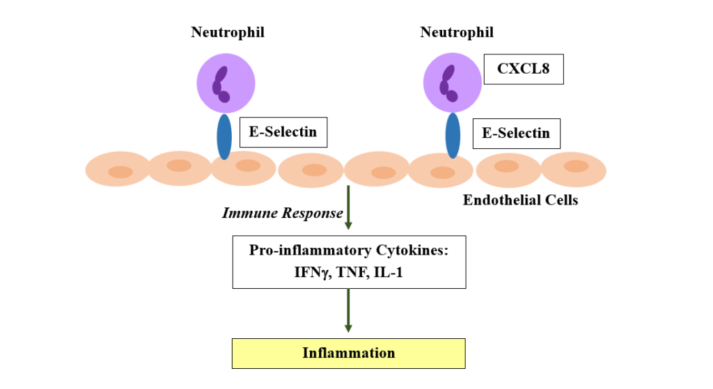
Fig. 2. Neuronal damage caused by viral infection can cause neuroinflammation.
CONCLUSIONS
Virus-infected cells in the brain can activate an immune response in which chemokines intervene. Chemokines play a fundamental role in the immune response, coordinating leukocyte migration and the antiviral response in the CNS, but they can also contribute to neuroinflammation and neuronal damage. Their effect can therefore be protective or detrimental depending on the chemokine, the receptor involved, the cell type, and the stage of the disease, participating both in infection control and, in some cases, in repair and neuroprotection.
Conflict of interest
The authors declare that they have no conflict of interest.
REFERENCES
- Jin T, Xu X, Hereld D. Chemotaxis, chemokine receptors and human disease. Cytokine. 2008;44(1):1-8. doi:10.1016/j.cyto.2008.06.017
- Kufareva I, Gustavsson M, Zheng Y, Stephens BS, Handel TM. What Do Structures Tell Us About Chemokine Receptor Function and Antagonism? Annual Review of Biophysics. 2017;46(1):175-198. doi:10.1146/annurev-biophys-051013-022942
- Hosking MP, Lane TE. The Role of Chemokines during Viral Infection of the CNS. , ed.Madhani HD. PLoS Pathogens. 2010;6(7):e1000937. doi:10.1371/journal.ppat.1000937
- Wu Y, Yoder A. Chemokine Coreceptor Signaling in HIV-1 Infection and Pathogenesis. , ed.Manchester M. PLoS Pathogens. 2009;5(12):e1000520. doi:10.1371/journal.ppat.1000520
- Dixit N, Simon SI. Chemokines, selectins and intracellular calcium flux: temporal and spatial cues for leukocyte arrest. Frontiers in Immunology. 2012;3. doi:10.3389/fimmu.2012.00188
- Laudanna C, Kim JY, Constantin G, Butcher EC. Rapid leukocyte integrin activation by chemokines. Immunological Reviews. 2002;186(1):37-46. doi:10.1034/j.1600-065x.2002.18604.x
- Bitoux MA, Stamenkovic I. Tumor-host interactions: the role of inflammation. Histochemistry and Cell Biology. 2008;130(6):1079-1090. doi:10.1007/s00418-008-0527-3
- Carnevale S, Di Ceglie I, Lasaracina AP, Molisso P, Jaillon S. Myeloid cells as sources and targets of IL-1 family cytokines in cancer. Seminars in Immunology. 2026;83:102048. doi:10.1016/j.smim.2026.102048
- Melchjorsen J, Sørensen LN, Paludan SR. Expression and function of chemokines during viral infections: from molecular mechanisms to in vivo function. Journal of Leukocyte Biology. 2003;74(3):331-343. doi:10.1189/jlb.1102577
- Chan YK, Gack MU. Viral evasion of intracellular DNA and RNA sensing. Nature Reviews Microbiology. 2016;14(6):360-373. doi:10.1038/nrmicro.2016.45
- Tan X, Sun L, Chen J, Chen ZJ. Detection of Microbial Infections Through Innate Immune Sensing of Nucleic Acids. Annual Review of Microbiology. 2018;72(1):447-478. doi:10.1146/annurev-micro-102215-095605
- Mahalingam S, Friedland JS, Heise MT, Rulli NE, Meanger J, Lidbury BA. Chemokines and viruses: friends or foes? Trends in Microbiology. 2003;11(8):383-391. doi:10.1016/s0966-842x(03)00157-4
- Lusso P. Chemokines and Viruses: The Dearest Enemies. Virology. 2000;273(2):228-240. doi:10.1006/viro.2000.0443
- Barry M, Bleackley RC. Cytotoxic T lymphocytes: all roads lead to death. Nature Reviews Immunology. 2002;2(6):401-409. doi:10.1038/nri819
- Hickman HD, Reynoso GV, Ngudiankama BF, et al. CXCR3 Chemokine Receptor Enables Local CD8+ T Cell Migration for the Destruction of Virus-Infected Cells. Immunity. 2015;42(3):524-537. doi:10.1016/j.immuni.2015.02.009
- Del-Val M, López D. Multiple proteases process viral antigens for presentation by MHC class I molecules to CD8+ T lymphocytes. Molecular Immunology. 2002;39(3-4):235-247. doi:10.1016/s0161-5890(02)00104-9
- Hansen TH, Bouvier M. MHC class I antigen presentation: learning from viral evasion strategies. Nature Reviews Immunology. 2009;9(7):503-513. doi:10.1038/nri2575
- Parry RV, Riley JL, Ward SG. Signalling to suit function: tailoring phosphoinositide 3-kinase during T-cell activation. Trends in Immunology. 2007;28(4):161-168. doi:10.1016/j.it.2007.02.004
- Agarwal P, Raghavan A, Nandiwada SL, et al. Gene Regulation and Chromatin Remodeling by IL-12 and Type I IFN in Programming for CD8 T Cell Effector Function and Memory. The Journal of Immunology. 2009;183(3):1695-1704. doi:10.4049/jimmunol.0900592
- Spolski R, Li P, Leonard WJ. Biology and regulation of IL-2: from molecular mechanisms to human therapy. Nature Reviews Immunology. 2018;18(10):648-659. doi:10.1038/s41577-018-0046-y
- Russell JH, Ley TJ. Lymphocyte-mediated cytotoxicity. Annual Review of Immunology. 2002;20(1):323-370. doi:10.1146/annurev.immunol.20.100201.131730
- Michlmayr D, McKimmie CS, Pingen M, et al. Defining the chemokine basis for leukocyte recruitment during viral encephalitis. Journal of Virology. 2014;88(17):9553-9567. doi:10.1128/JVI.03421-13
- Zhao J, Li Q, Ouyang X, et al. The effect of CX3CL1/ CX3CR1 signal axis on microglia in central nervous system diseases. Journal of Neurorestoratology. 2023;11(1):100042. doi:10.1016/j.jnrt.2023.100042
- Limatola C, Ransohoff RM. Modulating neurotoxicity through CX3CL1/CX3CR1 signaling. Frontiers in Cellular Neuroscience. 2014;8. doi:10.3389/fncel.2014.00229
- Roberts TK, Eugenin EA, Lopez L, et al. CCL2 disrupts the adherens junction: implications for neuroinflammation. Laboratory Investigation. 2012;92(8):1213-1233. doi:10.1038/labinvest.2012.80
- Mahad D, Callahan MK, Williams KA, et al. Modulating CCR2 and CCL2 at the blood–brain barrier: relevance for multiple sclerosis pathogenesis. Brain. 2005;129(1):212-223. doi:10.1093/brain/awh655
